
Total Joint Replacement: Hip Joint
The hip joint is a ball-and-socket joint that allows movement in many directions. It is involved in normal walking, running, bending, twisting, adduction and abduction of the legs.
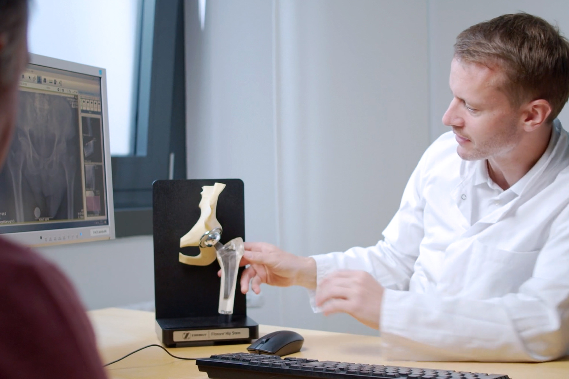
The hip joint
- Hip joint socket (acetabulum)
- Femoral head (spherical joint head)
- Femoral neck
- Pelvic bone
- Thigh bone (femur)
Non-surgical treatment: Hips
When patients with (more or less) severe hip ailments come to my practice, my first goal is to alleviate the pain with non-surgical treatment.
Based on the arthritic changes in X-ray images and the duration and severity of your ailments, we are well equipped to make a decision about which treatments are helpful.
I have had good experiences with a combination of several methods aimed at alleviating your pain and restoring the mobility of your hips.
These include
- natural and pharmacological inflammation inhibitors
- an analysis of your activities and, if necessary, a modification of your movement habits
- physiotherapy exercises that you perform yourself at home
- specific physiotherapy
- injections with cortisone, PRP (a form of plasma therapy) or hyaluronic acid.
You receive the injections right there on the spot during your appointment at my practice – this provides rapid pain relief and saves you time and inconvenience.
Throughout the entire therapeutic treatment, my team and I carefully supervise the progression of your cartilage damage.
Only once non-surgical treatment fails and the desired success is not achieved do we consider hip surgery.
Together we weigh up the options and make the – not always easy – decision of when surgery should be carried out and what type of hip implant is most suitable for you.
Surgical treatment: Hip surgery
During hip replacement surgery, the diseased hip joint is replaced with an artificial implant (total hip replacement or THR for short).
If you do require surgery, we take your individual anatomy and risks into account, as well as your desired activities and workload. In addition, we also discuss realistic expectations of the artificial joint.
What does hip surgery look like?
Before any operation, we discuss the procedure in detail and I answer all your questions so that you are well informed and feel confident about having the procedure carried out without fear.
The entire procedure usually takes an hour on average. Before this, you receive a general anaesthetic or a spinal anaesthetic (also known as a spinal block).
And then follows the hip surgery:
- Depending on your individual diagnosis, for the surgery I select either an approach in the anterior (front), anterolateral area of the hip joint (ALMIS method), a purely lateral (sideways) approach to the hip or a posterior (back) approach.
- Your natural hip socket in the pelvis is replaced with a prosthetic cup with or without bone cement.
- After this, we prepare the medullary cavity of the femur in order to precisely fix the prosthesis stem with or without bone cement.
- A spherical head is placed on the prosthesis stem. It establishes the mobile connection between the stem and the artificial hip socket.
- I use a trial implant to check the optimum mobility of the joint before inserting the original implant.
Finally, we close up the wound and produce an initial X-ray image following the operation.
After the hip surgery
During your inpatient stay, you will relearn how to walk confidently and independently with the help of crutches – generally without any limiting strain. You will be discharged once the conditions of your wounds and soft tissue have healed regularly, you have learned how to walk with confidence and there are no medical reasons opposing your discharge.
You can train yourself to walk without the crutches in your home environment. As a general rule, you should be able to drive independently again after 4 weeks . If you wish, we can also initiate rehab therapy. If the procedure is straightforward, however, this is not absolutely necessary; in this case, outpatient physiotherapy is sufficient. The first follow-up appointments take place 6 and 12 weeks after the surgery.
Hip joint revision surgery
An implanted prosthesis does not last forever; usually between 20 to 25 years.
Over the years, abrasion particles lead to an inflammatory reaction. This causes bone mass to degrade, as a result of which the prosthetic is "loosened" from the bone. Both the prosthetic cup as well as the stem can become loose separately from one another. Alternatively, both parts may also be affected by the loosening.
In any case, prompt action must be taken:
I place enormous value on finding out whether bacteria are involved before every revision surgery.
Revision surgery with bacteria present (2 surgery appointments)
If the loosening can be traced back to inflammation caused by bacteria, an infection is present. In this case, alongside the implant loosening, the infection (osteomyelitis) in particular must also be treated. To do so, the entire prosthesis must be removed, regardless of its degree of loosening. The prosthesis is then replaced with a placeholder containing antibiotics, and a 6-week treatment course of antibiotics takes place. Following this, what’s known as a revision endoprosthesis is implanted.
Revision surgery at an appointment
If there are no bacteria present, the loosening can only be traced back to an inflammatory reaction due to the abrasion. In this case, the replacement can take place in one surgery appointment and can be made conditional on the degree of loosening of the components. In this process, it is always only the loose part that is replaced.
Just as with the primary implants, I place value on careful surgical planning to ensure that I am able to react to extraordinary circumstances in the revision situation during the hip revision surgery – such as major bone defects, poor bone quality, fractures around the implant site. (The implant site is the area that will receive the implant.) This is why we keep a large repertoire of revision and special implants on hand or have ordered them in advance for the revision situation.
This allows us, the surgical team, to perform the optimum reconstruction of the biological situation of the joint.
During an extensive medical history interview, we find the right non-surgical or surgical therapy steps for you.
FAQ
What is the ALMIS method? What does an anterolateral approach to the hip mean?
To speed up rehabilitation and shorten your stay at the hospital, for the majority of hip surgeries I select the anterolateral approach to the hip joint in the front side area of the hip joint. With around 200 surgeries per year using the so-called ALMIS method, I consider this method to be advantageous as the muscle is not separated from the pelvis or the femur. The most important muscles for the functioning of the hip, the gluteal muscles, which are attached to the posterior and lateral pelvis and femur, are not affected.
This method of surgery is particularly interesting for patients who would like to opt for the path of "fast-track surgery" or who want to get back to work as soon as possible. They are able to start driving again, for example, as soon as they feel well and are no longer taking opioid painkillers.
In the event of pronounced deformities, previous surgeries or larger revision procedures, on the other hand, it may be necessary to go with a lateral approach.
What does a hip endoprosthesis look like?
The implant consists of the hip socket and the hip stem, onto which is placed a spherical head that moves in the socket. The joint components – i.e. the articulating surfaces that have direct contact with each other (gliding joint pairs) – are a ceramic or metal head that moves within a cup insert made of durable plastic (polyethylene) or ceramic.
All the materials used are specially designed for medical purposes. They are characterized by high abrasion resistance and tissue compatibility and enable pain-free and permanent function.
How is the new joint anchored in the bone?
- With the cement-free implant, the hip stem is pressed into the bone. The hip socket is similarly pressed or screwed in. Thanks to the bone-friendly material, these components heal rapidly as the bone grows into them and the joint is fixed in place in the long term.
- With the cemented endoprosthesis, the hip stem and socket are affixed in the pelvis and femur with a quick-hardening plastic known as bone cement.
The hybrid implant is a mixed method: with this implantation technique, one of the components (hip socket or stem) is attached without cement, and the other is anchored using cement.
What is the right implant for me?
A hip joint that has been destroyed by arthrosis always has to be fully replaced. This entails replacement with an artificial socket that is fitted into the pelvis, as well as a hip stem that is implanted into the femur and a spherical head that is placed on the hip stem and moves within the socket. Each type of implant features a wide variety of models in various sizes. The choice of implant is based on the bone quality of the patient. Using the data from the examination and a special X-ray image, I plan the hip surgery and determine the size, position and type of fixation of the implant. In this process, I also take any potential difference in leg length into account.
How long do artificial hip joints last?
Experience shows that artificial joints last 20 to 25 years. Deciding factors in the lifespan and functionality are the material of the gliding joint pairs, the physical demands and the bone quality.
How does osteoarthtritis of the hip progress?
Osteoarthritis of the hip joint is usually age-related wear and tear that involves abrasion of the cartilage. The abrasion particles cause an inflammatory reaction that leads to the increased formation of synovial fluid.
What is more, after it reaches a certain level, the inflammation itself has a destructive effect on the cartilage, leading to even more cartilage degradation.
The bone initially reacts to the increasing strain with intensified calcification (sclerosing). Later on, it becomes involved in the self-destructive inflammatory reaction: the bone is degraded and cysts form. To compensate for this, the body forms bony growths (osteophytes) at the edge of the joint to stiffen the destroyed joint in the long run and thus end the sensation of pain.
The disease follows a wave-like pattern:
it is entirely possible for incredibly strong feelings of pain to be followed by intervals in which patients are free from pain. As the disease progresses, however, the frequency of the episodes of pain increases to the same extent as the duration of the pain-free intervals decreases.
The most important message in this context is that osteoarthritis is not curable , but the progression and symptoms can be alleviated (see non-surgical treatment).
When the disease has progressed so far that physical therapy measures, painkillers and anti-inflammatory medication no longer have any effect, the only measure left is an total joint replacement (endoprosthesis).
Osteoarthritis develops, therefore, over various stages, which are easily visible and can be well assessed on X-ray images, which is why X-ray imaging is the most important diagnostic tool for arthrosis.